LAB 2 Report written by YU TIAM MENG
Name: Yu Tiam Meng
Matric no: 111437
2.1 Ocular Micrometer
Introduction
Ocular
micrometer is use in order to measure and compare the size of prokaryotic and
eukaryotic microorganisms. Microorganisms are measured with an ocular
micrometer which is inserted into the one of the microscope eyepieces. The
micrometer, which serves as a scale or rule, is a flat circle of glass upon which
are etched equally spaced divisions. This is not calibrated, and may be used at
several magnifications. When placed in the eyepiece, the line superimposed
certain distance markers on the microscope field. The actual distance
superimposed may be calibrated using a stage micrometer on which parallel lines
exactly 10µm
apart etched. By determining how many units of the ocular micrometer
superimpose a known distance on the stage micrometer, you can calculate the
exact distance each ocular division measures on the microscopic field. When you
change objectives you must recalibrate the system. After calibration of the
ocular micrometer, the stage micrometer is replaced with a slide containing
microorganisms. The dimensions of the cells may then be determined.
Objective
To measure and count cells using a microscope
Results

One of the observation
400x magnification
|
1000x magnification
|
||
Lactobacillus
|
Yeast
|
Lactobacillus
|
Yeast
|
Cells are too small and we can't observe under this magnification.
|
5 µm (2 divisions)
|
2 µm (2 divisions)
|
5 µm (5 divisions)
|
Discussion
- Steps in calculating the Ocular micrometer divisions with the use of different power objectives:
400x magnification
|
1000x magnification
|
||
Stage scale
|
Divisions in Ocular micrometer
|
Stage scale
|
Divisions in Ocular micrometer
|
0.05 mm
(we use 5 divisions of stage scale here because we can't see 1 divisions of stage scale clearly in 400x magnification)
|
20
|
0.01 mm
|
10
|
1 division in stage scale = 0.01 mm
At last we get,
400x magnification
|
1000x magnification
|
||
Stage scale
|
Divisions in Ocular micrometer
|
Stage scale
|
Divisions in Ocular micrometer
|
0.0025 mm
|
1 division
|
0.001 mm
|
1 division
|
2.5 µm
|
1 division
|
1 µm
|
1 division
|
- The actual size of microorganisms:
- The actual size of Lactobacillus = 2.0 to 4.0 micrometers in length
- The actual size of Yeast = typically 3–4 µm in diameter, although some yeasts can reach over 40 µm
Micrometry is the measurment of microorganisms. Since
microorganisms can be seen only under a microscope, suitable scale for their
measurements should be somewhere in the microscope itself. Ocular micrometer is
simply a disc of glass upon which are etched lines. When placed in the eye
piece, the ruled lines superimpose certain distance markers on the microscope
field. However, the scale on ocular micrometer does not have any standard
value. We can find out the value of one division of this unknown scale by calibrating
it with known scale. Thus actual value of one division of ocular micrometer is
found by using another known scale, the stage micrometer.First, look through
your microscope's eyepieces and determine whether there is an ocular micrometer
in place. Ocular micrometers appear as a scale of parallel black lines similar
to lines on a ruler, often with numbers indicating sequential measures of ten
lines.Calibrate the ocular micrometer if this has not been done previously.
Place a stage micrometer slide on the stage and view it through the eyepieces,
making sure that both eyepieces are focused. By rotating the eyepiece
containing the ocular micrometer and moving the stage micrometer slide, align
the two micrometers.The stage micrometer has divisions of known
dimensions; use these dimensions to determine the ocular micrometer dimensions
for the objective, or microscope lens, directly over the stage. For example, if
each stage micrometer division using a certain objective can be aligned with
ten ocular micrometer divisions, then each ocular micrometer division is
one-tenth of the known stage micrometer division length. In the example, if
each stage micrometer division measures 100 microns, then each ocular micrometer
division using the objective measures 10 microns.After calibrating the ocular
micrometer for one objective, repeat the procedure for the other microscope
objectives for greatest accuracy. Alternatively, the calibration of the other
objectives can be calculated from the measured objective calibration; however,
this method can lead to error because of variations in exact magnification. For
example, a division measuring 10 microns under the 10 times magnification
objective would be calculated to measure one micron under the 100 times
magnification objective. Place a slide on the microscope stage. Align the
ocular micrometer with the surface of the object on the slide to be measured by
rotating the eyepiece containing the micrometer and moving the microscope slide
with the object until the micrometer is aligned with the surface. Count the
number of micrometer divisions aligned along the surface.
Calculate the surface length by multiplying the number of measured micrometer divisions by the conversion factor determined through ocular micrometer calibration in step one. For example, if each division is one micrometer and the surface measured aligns with 10 divisions, then the surface measurement is 10 micrometers.
Calculate the surface length by multiplying the number of measured micrometer divisions by the conversion factor determined through ocular micrometer calibration in step one. For example, if each division is one micrometer and the surface measured aligns with 10 divisions, then the surface measurement is 10 micrometers.
Conclusion :
-Inaccurate measurements will result from not calibrating the reticule properly.-Because the micrometer measures a flat dimension, consider the three-dimensional aspect of the measured object. For example, the length of a curved surface may be longer than the ocular micrometer measured length.
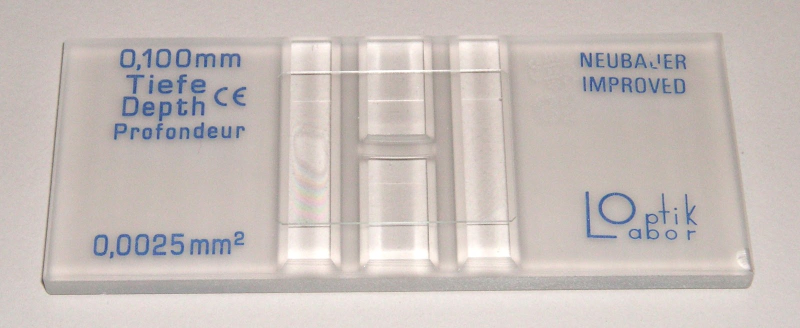
2.2 Neubauer Chamber
Introduction
Neubauer chambers are more convenient for counting microbes. The Neubauer is a heavy glass slide with two counting areas separated by a H-shaped trough. A special coverslip is placed over the counting arreas and sits a precise distance above them.
Objective
To measure and count cells using a microscope
Results

One of the observation
Average of cells' number = (31+34+35+36+34+35+41+40+35+37) / 10
= 35.8
Volume of small square = 0.2 x 0.2 x 0.1
= 4.0 x 10⁻³ mm3
= 4.0 x 10⁻⁶ cm3
= 4.0 x 10⁻⁶ mL
Average number of cells in one small
square :
(35.8 x 250,000) = 4 x 10⁻⁶
mL
2.2375 x 10¹²
cells = 1 mL
2.2375 x 10¹²
cells/mL
Discussions
hemocytometer.

Line Grid of the hemocytometer.
One can often determine cell density of a suspension spectrophotometrically, however that form of determination does not allow an assessment of cell viability, nor can one distinguish cell types. To prepare the counting chamber the mirror-like polished surface is carefully cleaned with lens paper. The coverslip is also cleaned. Coverslips for counting chambers are specially made and are thicker than those for conventional microscopy, since they must be heavy enough to overcome the surface tension of a drop of liquid. The coverslip is placed over the counting surface prior to putting on the cell suspension. The suspension is introduced into one of the V-shaped wells with a pasteur or other type of pipet. The area under the coverslip fills by capillary action. Enough liquid should be introduced so that the mirrored surface is just covered. The charged counting chamber is then placed on the microscope stage and the counting grid is brought into focus at low power.
It is essential to be extremely
careful with higher power objectives, since the counting chamber is much
thicker than a conventional slide. The chamber or an objective lens may be
damaged if the user is not not careful. One entire grid on standard
hemacytometers with Neubauer rulings can be seen at 40x (4x objective). The
main divisions separate the grid into 9 large squares (like a tic-tac-toe
grid). Each square has a surface area of one square mm, and the depth of the
chamber is 0.1 mm. Thus the entire counting grid lies under a volume of 0.9
mm-cubed.
Suspensions should be dilute enough
so that the cells or other particles do not overlap each other on the grid, and
should be uniformly distributed. To perform the count, determine the
magnification needed to recognize the desired cell type. Now systematically
count the cells in selected squares so that the total count is 100 cells or so
(number of cells needed for a statistically significant count). For large cells
this may mean counting the four large corner squares and the middle one. For a
dense suspension of small cells you may wish to count the cells in the four
1/25 sq. mm corners plus the middle square in the central square. Always decide
on a specific counting patter to avoid bias. For cells that overlap a ruling,
count a cell as "in" if it overlaps the top or right ruling, and
"out" if it overlaps the bottom or left ruling.
Here is a way to determine a
particle count using a Neubauer hemocytometer. Suppose that you conduct a count
as described above, and count 187 particles in the five small squares
described. Each square has an area of 1/25 mm-squared (that is, 0.04
mm-squared) and depth of 0.1 mm. The total volume in each square is
(0.04)x(0.1) = 0.004 mm-cubed. You have five squares with combined volume of
5x(0.004) = 0.02 mm-cubed. Thus you counted 187 particles in a volume of 0.02
mm-cubed, giving you 187/(0.02) = 9350 particles per mm-cubed. There are 1000
cubic millimeters in one cubic centimeter (same as a milliliter), so your
particle count is 9,350,000 per ml.
Cells are often large enough to
require counting over a larger surface area. For example, you might count the
total number of cells in the four large corner squares plus the middle
combined. Each square has surface area of 1 mm-squared and a depth of 0.1 mm,
giving it a volume of 0.1 mm-cubed. Suppose that you counted 125 cells (total)
in the five squares. We then have 125 cells per 0.5 mm-cubed, which is 250
cells/mm-cubed. Again, multiply by 1000 to determine cell count per ml
(250,000).
Sometimes we will need to dilute a
cell suspension to get the cell density low enough for counting. In that case we
will need to multiply final count by the
dilution factor. For example, suppose that for counting you had to dilute a
suspension of Chlamydomonas 10 fold. Suppose we obtained a final count of
250,000 cells/ml as described above. Then the count in the original (undiluted)
suspension is 10 x 250,000 which is 2,500,000 cells/ml.
Conclusion:
- Type of counting chambers: There are different types of counting chambers available, with different grid sizes. One counting chamber also has grids of different sizes. Take care that that you know the grid size and height (read the instruction manual) otherwise you’ll make calculation errors.
- Use the provided cover glasses: They are thicker than the standard 0.15mm cover glasses. They are therefore less flexible and the surface tension of the fluid will not deform them. This way the height of the fluid is standardized.
- Moving cells: Moving cells (such as sperm cells) are difficult to count. These cells must first be immobilized.
- Objective The hemocytometer is much thicker than a regular slide. Be careful that you do not crash the objective into the hemocytometer when focusing.
References:
LAB 2 Report written by Muhammad Aizat
Name: Muhammad Aizat b Mat Saad
Matric no: 111385
Matric no: 111385
LAB 2: MEASUREMENT AND COUNTING OF
CELLS USING MICROSCOPE
INTRODUCTION
2.1 Ocular
Micrometer
An ocular micrometer is a glass disk that attaches to a
microscope's eyepiece. An ocular micrometer has a ruler that allows the user to
measure the size of magnified objects. The distance between the marks on the
ruler depends upon the degree of magnification. The ruler on a typical ocular
micrometer has between 50 to 100 individual marks, is 2 mm long and has a distance
of 0.01 mm between marks.

 |
| Ocular micrometer |
Ocular micrometer is used in order to measure and compare the
size of prokaryotic and eukaryotic microorganisms. Microorganisms are measured
with an ocular micrometer which is inserted into the one of the microscope
eyepiece. The micrometer, which serves as scale or rule, is flat circle of
glass upon which are etched equally spaced divisions. This is not calibrated,
and may be used at several magnifications. When placed in the eyepiece, the
line superimposed certain distance markers on the microscope field. The actual
distance 10µm apart etched. By determining how many units of the ocular
micrometer superimposed a known
distance on stage micrometer, you can calculate the exact distance each ocular
division measures on the microscopic field. When you change objectives you must
recalibrate the system. After calibration of the ocular micrometer, the stage
micrometer is replaced with a slide containing microorganisms. The dimensions
of the cells may then be determined.
2.2 NEUBAUER CHAMBER
For microbiology, cell culture, and many applications that
require use of suspensions of cells it is necessary to determine cell
concentration. One can often determine cell density of a suspension
spectrophotometrically, however that form of determination does not allow an
assessment of cell viability, nor can one distinguish cell types. A device used
for determining the number of cells per unit volume of a suspension is called a
counting chamber. The most widely used type of chamber is called a Neubauer
chamber, since it was originally designed for performing blood cell counts.
Neubauer chamber are more convenient for counting microbes. The neubauer
chamber is a heavy glass slide with two counting areas separated by a H-shaped
through figure. To prepare the counting chamber the mirror-like polished
surface is carefully cleaned with lens paper. The coverslip is also cleaned.
Coverslips for counting chambers are specially made and are thicker than those
for conventional microscopy, since they must be heavy enough to overcome the
surface tension of a drop of liquid. The coverslip is placed over the counting
surface prior to putting on the cell suspension. The suspension is introduced
into one of the V-shaped wells with a pasteur or other type of pipet. The area
under the coverslip fills by capillary action. Enough liquid should be introduced
so that the mirrored surface is just covered. The charged counting chamber is
then placed on the microscope stage and the counting grid is brought into focus
at low power.


OBEJCTIVE
To measure
and count cell using a microscope
RESULT
2.1 Ocular Micrometer

Lactobacillus - 1000x
magnification
Size= 2 µm (2 divisions)

Yeast
400x magnification
|
1000x magnification
|
Size= 5 µm (2
divisions)
|
Size= 5 µm (5
divisions))
|
2.2 NEUBAUER CHAMBER
100 x 10
magnification (oil-immersion).
Calculation
:
Average of
cell :
= (31 + 34 +
35 + 36 + 34 + 35 + 41 + 40 + 35 + 37 ) ÷ 10
= 35.8
Volume of
square :
0.2 x 0.2 x 0.1= 4 x 10 ⁻ᶟ mmᶟ
4 x 10 ⁻ᶟ mmᶟ
x 10 ⁻ᶟ = 4 x 10⁻⁶ cmᶟ
4 x 10⁻⁶ cmᶟ
= 4 x 10⁻⁶ ml
Concentration
of cell :
= 35.8 ÷ ( 4
x 10-6 )
= 8.95 x 10 ⁶
cells/ml
DISCUSSION
2.1 Ocular Micrometer
Ocular micrometers
have no units on them , they are like a ruler with marks but no numbers. In
order to use one to measure something under a microscope, you must assign
numbers to the marks. This is done by looking
through your ocular micrometer at a stage micrometer mounted on a slide. The stage
micrometer is just a ruler with fixed known distances, so you can use it to
tell how far apart marks are on the ocular micrometer. This has to be done
because the marks on the ocular micrometer are different distances apart
depending on the magnification used on the microscope. It must be calibrated
for each objective.
1) How to use a ocular micrometer:
2) Attach the ocular micrometer to the
microscope eyepiece by unscrewing the eyepiece cap, placing the ocular
micrometer over the lens and screwing the eyepiece cap back into place.
3) Slide the stage micrometer onto the
microscope slide stage. Adjust the microscope to the lowest possible
magnification, which should bring the grid on the stage micrometer into focus.

4) Move the stage micrometer until the
measurement marks on the ocular micrometer align with the measurement marks on
the stage micrometer. The measurement "0" on the ocular micrometer
should line up with the measurement "0.0" on the stage micrometer.
5) Count the number of measurement marks
until the measurements of both the micrometers line up again. At 4x
magnification (the lowest setting on most microscopes), the two micrometers
will line up again at "3" on the ocular micrometer and
"0.3" on the stage micrometer.
6) Write down the number of measurement
marks between the aligning measurements for the two micrometers. The distance
between measurement marks is 0.01 mm, so you can now determine the distance
between coinciding measurement marks. Repeat the exercise at higher
magnifications (10x, 40x and 100x), and record these values as well.
Calculating
the ocular micrometer division scale on different magnifications( 400x and
1000x):
1) 400 x magnification
20 eyepiece divisions = 5 stage divisions
= 50 µm
1 eyepiece division = 50/20
= 2.5 µm
2) 1000 x magnification
10 eyepiece divisions = 1 stage divisions = 10 µm
1
eyepiece division = 10/10
= 1.0 µm
2.2 NEUBAUER CHAMBER
1) Preparing
the sample
The fluid
containing the cells must be appropriately prepared before applying it to the
hemocytometer.
·
Proper
mixing: The fluid should be a homogenous suspension. Cells that stick together
in clumps are difficult to count and they are not evenly distributed.
·
Appropriate
concentration: The concentration of the cells should neither be too high or too
low. If the concentration is too high, then the cells overlap and are difficult
to count. A low concentration of only a few cells per square results in a
higher statistical error and it is then necessary to count more squares (which
takes time). Suspensions that have a too high concentration should be diluted
1:10, 1:100 and 1:1000. A 1:10 dilution can be made by taking 1 part of the
sample and mixing it with 9 parts water. The dilution must later be considered
when calculating the final concentration.
2) Counting the cells

·
Counting
cells that are on a line: Cells that are on the line of a grid require special
attention. Cells that touch the top and right lines of a square should not be
counted, cells on the bottom and left side should be counted.
·
Number
of squares to count: The lower the concentration, the more squares should be
counted. Otherwise one introduces statistical errors. How many squares? To find
out one could calculate the cell concentration per ml based on the numbers
obtained from 2 different squares. If the final result is very different, then
this can be an indication of sampling error.
3) Calculating
the cell density
Here it is
necessary to do some simple math. The following numbers are needed: number of
cells counted in a square, area of the square, height of the sample, dilution
factor. The objective is to find the number of cells in 1ml of original
solution.
·
Step
1 – Averaging: need not count all of the cells in a large square (1mmx1mm), count
the cells in 10 selected small squares and find the average for one small
square. It is necessary to average the results first before proceeding.
·
Step
2 – Computing the volume: It is necessary to determine the volume represented
by the square. The width and height of the square (e.g. 0.25mm x 0.25mm) must
be multiplied by the height of the sample (often printed on the hemocytometer,
in this example it is 0.1mm): v = 0.25mm x 0.25mm x 0.1mm = 0.00625mm³ = 6.25 x
10 ⁻ᶟ mmᶟ
= 6.25 x 10 ⁻ᶟ x 10⁻ᶟ cmᶟ
= 6.25 x 10 ⁻⁶ cmᶟ
= 6.25 x 10 ⁻⁶ mL
·
Step
3 – Calculating the number of cells in 1 ml: if there are 123.456 cells 6.25 x
10 ⁻⁶ mL then how many cells are there in 1ml? We do simple direct proportion:
123.456cells/6.25 x 10 ⁻⁶ mL = X
Concentration of cell = 19 752 960 cells/ml
·
Step
4 – Correcting for dilution: If the sample was diluted before counting, then
this must be taking into consideration as well. We assume that the sample was
diluted 1:10. The final result is therefore 19 752 960 cells x 10 = 197 529 600
cells in 1 ml.
4)Precaution:
·
Type
of counting chambers: There are different types of counting chambers available,
with different grid sizes. One counting chamber also has grids of different
sizes. Take care that that you know the grid size and height (read the
instruction manual) otherwise you’ll make calculation errors.
·
Use
the provided cover glasses: They are thicker than the standard 0.15mm cover
glasses. They are therefore less flexible and the surface tension of the fluid
will not deform them. This way the height of the fluid is standardized.
·
Moving
cells: Moving cells are difficult to count. These cells must first be
immobilized.
·
Objective
The hemocytometer is much thicker than a regular slide. Be careful that you do
not crash the objective into the hemocytometer when focusing.
CONCLUSIONS
2.1 Ocular Micrometer
Size of the
cell can be measure and the size between prokaryotic and eukaryotic can be
compare by using a microscope with the
ocular mirometer inserted into the eyepieces. The size of Lactobacillus(prokaryotic) is 2 µm (2
divisions), which are so small thus can only be measured with 1000x magnification.
While the size of Yeast(eukaryotic) is: 1) 5 µm (2 divisions) - 400x magnification 2) 5 µm (5 divisions)- 1000x magnification
2.2 NEUBAUER CHAMBER
The average
of the cell is 38.5 cells,the volume of one small box of the cells is 6.25 x 10
-⁶ mL and the concentration of cell is 8.95 x 10 ⁶ cells/ml.
References:
http://www.microbehunter.com/2010/06/27/the-hemocytometer-counting-chamber/
No comments:
Post a Comment