Before we continue our lab reports, we would like to introduce ourselves first.
Here we go, here are our group members

Hi, I'm Tiam Meng^^ !!!


Hi, I'm Azizul

Okay, let's begin with our lab reports.
Lab 1 Report Written by Yu Tiam Meng
Name : Yu Tiam Meng
Matrix number : 111437
LAB 1 : PRINCIPLES AND USE OF MICROSCOPE
1.1 Setting up and using the microscope
Introduction:
In order to be seen, microorganisms need to be magnified. Despite advances in other area of microscopy (for example, the electron microscopy), the light microscopy is still the instrument most frequently used for viewing microorganisms.
In order to be seen, microorganisms need to be magnified. Despite advances in other area of microscopy (for example, the electron microscopy), the light microscopy is still the instrument most frequently used for viewing microorganisms.
Objective:
Learn to use a simple bright-field microscope correctly.
Learn to use a simple bright-field microscope correctly.
Results:
Total magnification = objective lens power x eyepiece lens power(10x)
Total magnification = objective lens power x eyepiece lens power(10x)
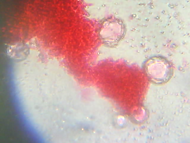
Diplococcus under 40x magnification:

Diplococcus under 100x magnification:

Diplococcus under 400x magnification:
Discussions:
1) The light intensity is adjusted to make sure that the pictures captured are clear and not too bright.
.jpg)
2) Colony morphology of Diplococcus: A diplococcus is a round bacterium that typically occurs in the form of two joined cells. Its name comes from diplo, meaning double, and coccus, meaning berry.
Conclusion:
I managed to get the view of specimen by adjusting the fine and coarse adjustment knob but I forgot to copy down the full name of specimen.
References:
1.2 Examination of cells
Introduction :
Because of their extreme minuteness, bacteria are not generally studied with the low-power or high-power dry objectives. Instead they are stained and observed with the oil immersion objective.
The wet mount methods enables you to study the sizes and shapesof living microorganisms (drying or staining microorganism distort them). It also enables you to determine if cells are motile. The wet mount method is quick and easy, and does not require special equipment.
-To provide an experience in the use of microscope.
-To illustrate the diversity of cells and microorganisms.
Result :
Under 100x objective X 10x eyepiece = 1000x magnification
1.Saccharomyces cerevisiae (Yeast)

2.Lactobacillus fermentum

Discussions:
1) Oil immersion is a technique used to increase the resolution of a microscope. This is achieved by immersing both the objective lens and the specimen in a transparent oil of high refractive index, thereby increasing the numerical aperture of the objective lens.
2)Colony morphology of Lactobacillus fermentum:
- They ferment sugars and produce lactic acid and they are aerotolerant anaerobes(they grow fermentatively with or without oxygen)
- They are in rod shape, tiny in size, smooth surface, and violet in colour.
3)Colony morphology of Saccharomyces cerivisiae:
- Yeast are nonfilamentous, unicellular fungi that are typically spherical or oval.
- Yeast are capable of facultative anearobic growth.
- Mycelium absent. Vegetative cells reproducing by multilateral budding, without a mucous coating. Asci morphologically similar to vegetative cells, not in well defined-chains, thin-walled, 1- to 4-spored, evanescent or semi-persistent, without an obvious discharge mechanism. Ascospores usually spherical, oftten ornamented with equatorial ridges. Fermentation positive, coenzyme usually Q-6.
.jpg)
.jpg)
.jpg)
Conclusion:
The use of immersion oil and 1000x magnification enable me to get a clearer image of specimens.
References:
1) http://en.wikipedia.org/wiki/Oil_immersion
2) MICROBIOLOGY (AN INTRODUCTION) FOURTH EDITION by Tortora , Funke , Case , 1992 by The Benjamin/Cummings Publishing Company, Inc.
3) Introduction to Microbiology (Second Edition) by John L. Ingraham/Catherine A. Ingraham, 2000 by Brooks/Cole.
3) Introduction to Microbiology (Second Edition) by John L. Ingraham/Catherine A. Ingraham, 2000 by Brooks/Cole.
LAB 1 REPORT
WRITTEN BY ISKANDAR
Introduction:
- microorganisms need
to be seen in a microscope. 1.1
Introduction to Microscope
Microscope is an instrument that is used to observed sample of
microorganisms under high magnification. Light microscope is type of microscope
that is frequently used for viewing microorganisms. A brightfield microscope is a type of
microscope that utilizes the brightfield microscopy technique. This is a
type of optical microscopy illumination that is the simplest and most reliable
among all the microscopy techniques out there. This microscopy technique
uses white light for illumination that comes from below the specimen in what is
called transmitted illumination.However, it can also be considered as the
method of lighting in a stereoscope using upper incident illumination.
Utilizing
the most basic type of microscopy, the bright field microscope is widely used
from simple and starting types like kid or child school microscope to
university or professional level models. Just as a simple compound light microscope
that uses brightfield microscopy is important to students as well as children,
bright field microscopes are also indispensable to biologists as well as
microbiologists in their respective fields.
There is 4 different magnification of objective lenses. That is 4x
magnification, 10x magnification, 40x magnification and 100x magnification. The
objective lens focuses the light passing through the specimen to form a
magnified primary image. There is also an eyepiece tube that receives light
coming through the objective lens and redirects it to the eyepiece.
Magnification of the eyepiece is 10x magnification. The eyepiece consists of
several lenses that collect the light, focus it and transmit it to the eye.
Total image of magnification is calculated by multiplying the objective lens
power multiplication by the eyepiece lens power multiplication.
The resolving power of resolution of microscope is its ability to
distinguish two very small and closely spaced objects as separate entities. A
number of factors affect resolution is the condenser diaphragm. Closing the
diaphragm image contrast but decreased the resolution, opening the diaphragm
decreases the contrast but increased the resolution.
Eyepiece or Ocular is what you look
through at the top of the microscope. Typically, standard eyepieces have a
magnifying power of 10x. Optional eyepieces of varying powers are available,
typically from 5x-30x.
Eyepiece tube holds the eyepieces in place
above the objective lens. Binocular microscope heads
typically incorporate a diopter adjustment ring that allows for the possible
inconsistencies of our eyesight in one or both eyes. The monocular (single eye usage)
microscope does not need a diopter. Binocular
microscopes also swivel (Interpupillary
Adjustment) to allow for different distances between the eyes of different
individuals.
Objective Lenses are the primary optical lenses
on a microscope. They range from 4x-100x and typically, include, three, four or
five on lens on most microscopes. Objectives can be forward or rear-facing.
Nosepiece houses the objectives. The
objectives are exposed and are mounted on a rotating turret so that different objectives
can be conveniently selected. Standard objectives include 4x, 10x, 40x and 100x
although different power objectives are available.
Coarse and Fine Focus knobs are used to focus the
microscope. Increasingly, they are coaxial knobs - that is to say they are
built on the same axis with the fine focus knob on the outside. Coaxial focus
knobs are more convenient since the viewer does not have to grope for a
different knob.
Stage is where the specimen to be
viewed is placed. A mechanical stage is used when working at higher
magnifications where delicate movements of the specimen slide are required.
Stage Clips are
used when there is no mechanical stage. The viewer is required to move the
slide manually to view different sections of the specimen.
Aperture is
the hole in the stage through which the base (transmitted) light reaches the
stage.
Illuminator is the light source for a microscope,
typically located in the base of the microscope. Most light microscopes use low
voltage, halogen bulbs with continuous variable lighting control located within
the base.
Condenser is used to collect and focus
the light from the illuminator on to the specimen. It is located under the
stage often in conjunction with an iris diaphragm.
Iris Diaphragm controls the amount of light
reaching the specimen. It is located above the condenser and below the stage.
Most high quality microscopes include an Abbe condenser with an iris diaphragm.
Combined, they control both the focus and quantity of light applied to the
specimen.
Condenser
Focus Knob moves the condenser up or down
to control the lighting focus on the specimen.
Objective:-
To learn a simple
bright field microscope correctly.
Material and reagents:
Microscope slide and
cover slip
Procedure:
Setting up:
Sit comfortably on
stool and knee under the bench and move the microscope to look to both
eyepieces without any strain.
Microscope is turned
on by using main switches.
By using the
brightness control, the light intensity is adjusted until position 5.
Nosepiece is rotated
until 4x objective lens is brought to the light path.
Clean slide with a
mark on it is placed on the stage and is secured with a spring clip. By using
coaxial stage control knobs, the slides is moved to the light path.
Both eyepieces is
adjusted until a single circle of light can be seen.
Tube length adjustment
(diopter) ring on right eyepiece was rotated until it match the interpupillary.
By using right eye
only, slide with the mark is adjusted with coarse and fine adjustment knobs
until it is focused.
By using left eye
only, slide with the mark is adjusted with coarse and fine adjustment knobs
until it is focused.
Low power (10x)
objective viewing:
Slide that is marked
with marker pen is replaced with specimen slide.
Stage is moved to
obtained view of specimen and focus is adjusted by using fine adjustment knob.
Then 10x objective lens is changed.
By placing an object
in the centre of the glass above light source, the condenser is focused by
adjusting the light condenser.
Object is removed out
of focused by lowering the condenser sufficiently.
Eyepiece is replaced
and re-focused with fine adjustment to obtained better image from specimen with
poor contrast.
High power (40x) objective
viewing :
Specimen was focused
with 10x objective lens. From side of microscope, 40x objective lens is
changed.
Condenser is raised
within 1cm of slide.
Specimen is focused by
using fine adjustment knob, light intensity is adjusted using brightness
control if necessary.
Condenser diaphragm is
adjusted for optimum contrast.
Oil immersion (100x)
objective viewing :
Specimen was focused
with 40x objective lens. From side of microscope, 100x objective lens is
changed and objective lens is not allow to touch the slide.
Objective lens is
carefully turned to one side and drops of oil is placed onto the slide.
Objective lens is turned again until it is in the light path.
Condenser is raised as
closed to the slide as possible.
Specimen is focused by
using fine adjustment knob, light intensity is adjusted using brightness
control if necessary.
Condenser diaphragm is
adjusted for optimum contrast.
After used :
Specimen slide is
removed and discarded into appropriate discard container.
Light brightness
control is reset to its lowest setting.
Lowest power objective
is reset to working position.
Oil from the 100x
objective lens is cleaned by using lens tissue.
Microscope is turned
off from the switch and power point, cord is disconnect and carefully wrapped
around base of microscope.
Cover is replaced.
Care of microscope :
Microscope is carried
by holding it firmly at the arm and support from the base. The instrument is
kept upright.
Never placed the
microscope at the edge of bench.
Only specified lens
tissue is used to clean the lenses.
Cover slip is used
when examining object or organism that is mounted with water or other fluids.
Stage was always
lowered when placing or removing slide.
Lowest power objective
lens was always placed in working position after the microscope had been used.
What do we observe is=
The following picture
is 1) sample with 4x10 magnification. 2) sample with 10x10 magnification 3)
sample with 40x10 magnification.
1.2 examination of cells
Introduction
Because of their extreme minuteness ,bacteria are not generally studied with the low power or high power dry objects. Instead they are stained and observed with the oil immersion objective.
The wet mount methods enables you to study the size and shapes of living microorganism. It also enables you to determine if the cell are motile. The wet mount method is quick and easy and does not require any special equipment.
Objective
- To provide an experience in the use of microscope.
- To illustrate the diversity of cells and microorganism.
Material and reagents
- Culture
- Immersion oil
- Lens tissue
- A microscope slide containing stained microorganism
- Inoculating loop
- Bunsen burner
- Slip and cover slip.
Procedure
Stained cells:
- The microscope is setted up as described and the slide is examined under the oil immersion lens
- The shape and size of the organism is observed. Picture are been captured.
The wet mount:
- A sterile pasteur pipette is used to aseptically transfer one drop of culture to the centre of the glass slide.
- A marker pen is used to mark the cover slip.
- The cover slip is taken and turned upside down so that the marker pen is faced down.
- The slide on the microscope stage is placed and 4x objective focus is used on the culture.
- The cell is observed using 10x and 40x objectives.
- The cells is observed by using oil immersion.
- Pictures has been captured.
- The process is repeated with other cultures.
Observation:
Stained cell:-

Stained cells slide 4x10 magnification

Stained cell 10x10 magnification

Stained cell 40x10 magnification
Wetted mount.

Saccharomyces cerevisea sp. Under 100x10 magnification under oil immersion.

Lactobacillus fermentum sp. Under 100x10 magnification under oil immersion.
Discussion:
Stained Cell
Bacteria are stain so that they can be more easily visualized under the microscope. Some stains can also be used to identify and classify bacteria. Gram stain is a method of staining bacteria using a dye called crystal (gentian) violet. Gram's method helps distinguish between different types of bacteria species into two large groups.
The gram-staining characteristics of bacteria are denoted as Gram-positive or Gram-negative, depending upon whether the bacteria take up and retain the crystal violet stain or not. The Gram stain is almost always the first step in the identification of a bacterial organism, and is the default stain performed by laboratories over a sample when no specific culture is referred. A Gram positive results in a purple/blue color while a Gram negative results in a pink/red color.
Gram-positive bacteria retain the color of the crystal violet stain in the Gram stain. This is characteristic of bacteria that have a cell wall composed of a thick layer of a particular substance, specificallypeptidologlycan containing teichoic and lipoteichoic acid complexed to the peptidoglycan. Molecules of crystal violet combine with iodine molecules to form a complex within the microbial cell. The addition of decolorizer effectively dehydrates the peptidoglycan layer preventing the extraction of the crystal violet–iodine complex from the interior of the cell. The final step consists of the addition of safranin counterstain. The color from the retained crystal violet-iodine complex obscures the safranin counterstain to give the cells a dark blue, or purple, appearance via high resolution light microscopy.
The Gram-positive bacteria include staphylococci ("staph"), streptococci ("strep"), pneumococci, and the bacterium responsible for diphtheria (Cornynebacteriumdiphtheriae) and anthrax (Bacillus anthracis).
Gram-negative bacteria lose the crystal violet stain (and take the color of the red counterstain) in Gram's method of staining. This is characteristic of bacteria that have a cell wall composed of a thin layer of a particular substance (specifically, peptidoglycan covered by an outer membrane of lipoprotein and lipopolysaccharide containing endotoxin). Molecules of crystal violet combine with iodine molecules to form a complex within the microbial cell. The addition of Gram decolorizer effectively extracts the outer membrane and dehydrates the peptidoglycan layer. It is presumed that the inner membrane is extracted by the Gram decolorizer, but remains associated with the mureinsacculus . However, the thinner dehydrated peptidoglycan layer cannot prevent the extraction of the crystal violet–iodine complex from the interior of the cell through the peptidoglycan layer. The final step consists of the addition of safranin counterstain. As the crystal violet-iodine complex has been extracted from the cells, the safranin counterstain gives the cells a pink, or red, appearance via high resolution light microscopy.
As for the lactobacillus fermentum, the shape is rod shape.mostly different length and the colour are transluscent. The elevation cannot be recognize and the size are punctiform. The surface of the bacteria mentioned are smooth and shiny.
As for the saccharomyces cerevisea the shape is almost circular and we can see the nucleus and some of the nucleus are undergoing mitosis and there is no elevation and the suface of the bacteria should be shinny and smooth.
References:
1) http://en.wikipedia.org/wiki/Oil_immersion
2) MICROBIOLOGY (AN INTRODUCTION) FOURTH EDITION by Tortora , Funke , Case , 1992 by The Benjamin/Cummings Publishing Company, Inc.
3) Introduction to Microbiology (Second Edition) by John L. Ingraham/Catherine A. Ingraham, 2000 by Brooks/Cole.
3) Introduction to Microbiology (Second Edition) by John L. Ingraham/Catherine A. Ingraham, 2000 by Brooks/Cole.
LAB 1 WRITTEN BY AZIZUL
Name : Ahmad Azizul Bin Md Sadik
Matrix No : 114116
LAB 1 : PRINCIPLES & USED OF MICROSCOPE
1.1 Introduction to Microscope
Microscope is an instrument that is used to observed sample of microorganisms under high magnification. Light microscope is type of microscope that is frequently used for viewing microorganisms. A brightfield microscope is a type of microscope that utilizes the brightfield microscopy technique. This is a type of optical microscopy illumination that is the simplest and most reliable among all the microscopy techniques out there. This microscopy technique uses white light for illumination that comes from below the specimen in what is called transmitted illumination.However, it can also be considered as the method of lighting in a stereoscope using upper incident illumination.
Utilizing the most basic type of microscopy, the bright field microscope is widely used from simple and starting types like kid or child school microscope to university or professional level models. Just as a simple compound light microscope that uses brightfield microscopy is important to students as well as children, bright field microscopes are also indispensable to biologists as well as microbiologists in their respective fields.
There is 4 different magnification of objective lenses. That is 4x magnification, 10x magnification, 40x magnification and 100x magnification. The objective lens focuses the light passing through the specimen to form a magnified primary image. There is also an eyepiece tube that receives light coming through the objective lens and redirects it to the eyepiece. Magnification of the eyepiece is 10x magnification. The eyepiece consists of several lenses that collect the light, focus it and transmit it to the eye. Total image of magnification is calculated by multiplying the objective lens power multiplication by the eyepiece lens power multiplication.

Eyepiece or Ocular is what you look through at the top of the microscope. Typically, standard eyepieces have a magnifying power of 10x. Optional eyepieces of varying powers are available, typically from 5x-30x.
Eyepiece tube holds the eyepieces in place above the objective lens. Binocular microscope heads typically incorporate a diopter adjustment ring that allows for the possible inconsistencies of our eyesight in one or both eyes. The monocular (single eye usage) microscope does not need a diopter. Binocular microscopes also swivel (Interpupillary Adjustment) to allow for different distances between the eyes of different individuals.
Objective Lenses are the primary optical lenses on a microscope. They range from 4x-100x and typically, include, three, four or five on lens on most microscopes. Objectives can be forward or rear-facing.
Nosepiece houses the objectives. The objectives are exposed and are mounted on a rotating turret so that different objectives can be conveniently selected. Standard objectives include 4x, 10x, 40x and 100x although different power objectives are available.
Coarse and Fine Focus knobs are used to focus the microscope. Increasingly, they are coaxial knobs - that is to say they are built on the same axis with the fine focus knob on the outside. Coaxial focus knobs are more convenient since the viewer does not have to grope for a different knob.
Stage is where the specimen to be viewed is placed. A mechanical stage is used when working at higher magnifications where delicate movements of the specimen slide are required.
Stage Clips are used when there is no mechanical stage. The viewer is required to move the slide manually to view different sections of the specimen.
Aperture is the hole in the stage through which the base (transmitted) light reaches the stage.
Illuminator is the light source for a microscope, typically located in the base of the microscope. Most light microscopes use low voltage, halogen bulbs with continuous variable lighting control located within the base.
Condenser is used to collect and focus the light from the illuminator on to the specimen. It is located under the stage often in conjunction with an iris diaphragm.
Iris Diaphragm controls the amount of light reaching the specimen. It is located above the condenser and below the stage. Most high quality microscopes include an Abbe condenser with an iris diaphragm. Combined, they control both the focus and quantity of light applied to the specimen.
1.2 Examination of Cells
Introduction
Historically, the study of cell biology could not have happened without the invention of microscopes because cells were not known to exist before Antonin van Leeuwenhoek and Robert Hooke saw them in their primitive microscopes Today, much cell biology research still requires careful microscopic examination of cells and their internal structures. It is not too strong a statement to say that microscopy is the single most important tool for the cell biologist.
Because of the extreme minuteness, bacteria are not generally studied with the low-power or high-power dry objectives. Instead it was stained and observed with the oil immersion objective.
The wet mount methods enables you to study the sizes and shapes of living microorganisms. It also enables to determine whether the cells are motile. The wet mount is quick and easy, also it does not require any special equipment.
Objectives :
To provide an experience in the use of microscope.
To illustrate the diversity of cells and microorganisms.
Materials and Reagents :
Culture
Immersion oil
Lens tissue
Microscope slide containing microorganisms
Inoculating loop
Bunsen burner
Slide and coverslip
Observations :

Observation under 4x10 magnification.

Observation under 10x10 magnification.

Observation under 40x10 magnification.
Sample for wet mount :

Observation under 100x10 magnification (oil immersion) for yeast sample, Saccharomyces cerevisiae.

Observation under 100x10 magnification (oil immersion) for Lactobacillus fermentum.
Discussion :
Gram staining is a common technique used to differentiate
two large groups of bacteria based on their different cell wall constituents.
The Gram stain procedure distinguishes between Gram positive and Gram negative
groups by coloring these cells red or violet. Gram positive bacteria stain
violet due to the presence of a thick layer of peptidoglycan in their cell
walls, which retains the crystal violet these cells are stained with.
Alternatively, Gram negative bacteria stain red, which is attributed to a
thinner peptidoglycan wall, which does not retain the crystal violet during the
decoloring process.
The Gram stain also used to detect the presence
of bacteria, yeast, and other cells in direct smears prepared from swabs,
aspirates, secretions, etc. from any part of the body where infection is suspected. Direct smears are often
made of throat swabs, sputum, genital swabs, wounds, abscesses, cerebrospinal
fluid (CSF), serous fluids, joint fluid, urine, and stool. Gram stain is also
performed to help identify colonies isolated from cultures. In addition to
gram-negative or gram-positive, organisms are evaluated for size, shape,
arrangement, number, and any special characteristics such as bipolar staining
and the presence of spores. These characteristics often point the way to the
most efficient selection of biochemical tests needed to identify the organism.
The finding of organisms on direct examination of some specimens is sufficient
to establish a preliminary diagnosis and justify immediate antibiotic treatment
pending confirmation by culture or other means. The Gram stain is very useful
in identifying anaerobic bacteria by comparing the microscopic morphology and
number of organisms to culture results. Significant numbers of characteristic
bacteria on Gram stain not appearing on aerobic culture often signals the
presence of an anaerobic infection.
Staphylococcus aureus forms a fairly large yellow colony on rich medium. S.
aureus is often hemolytic
on blood agar. Staphylococci are facultative anaerobes that grow by aerobic
respiration or by fermentation that yields principally lactic acid. The
bacteria are catalase-positive and oxidase-negative. S. aureus can grow at a temperature range of 15
to 45 degrees and at NaCl concentrations as high as 15 percent. S.
aureus should always be considered
a potential pathogen. Staphylococci are perfectly spherical cells about 1
micrometer in diameter. The staphylococci grow in clusters because the cells
divide successively in three perpendicular planes with the sister cells remaining
attached to one another following each successive division. Since the exact
point of attachment of sister cells may not be within the divisional plane, and
the cells may change position slightly while remaining attached, the result is
formation of an irregular cluster of cells.
The shape and configuration of the Gram-positive cocci helps to distinguish staphylococci from streptococci. Streptococci are slightly oblong cells that usually grow in chains because they divide in one plane only, similar to a bacillus. Without a microscope, the catalase test is important in distinguishing streptococci (catalase-negative) from staphylococci, which are vigorous catalase-producers. The test is performed by adding 3% hydrogen peroxide to a colony on an agar plate or slant. Catalase-positive cultures produce O2 and bubble at once. The test should not be done on blood agar because blood itself contains catalase.
The shape and configuration of the Gram-positive cocci helps to distinguish staphylococci from streptococci. Streptococci are slightly oblong cells that usually grow in chains because they divide in one plane only, similar to a bacillus. Without a microscope, the catalase test is important in distinguishing streptococci (catalase-negative) from staphylococci, which are vigorous catalase-producers. The test is performed by adding 3% hydrogen peroxide to a colony on an agar plate or slant. Catalase-positive cultures produce O2 and bubble at once. The test should not be done on blood agar because blood itself contains catalase.
Lactobacillus
fermentum is a Gram-positive species of bacterium in
the genus Lactobacillus. It is associated with active dental caries lesions. It is also commonly found in
fermenting animal and plant material. It
has been found in sourdough. A few strains are considered probiotic or "friendly"
bacteria in animals and at least
one strain has been applied to treat urogenital infections in women. Lactobacillus fermentum belongs to the genus Lactobacillus. Species in this genus are used for a wide variety of
applications. These applications include food and feed fermentation. It has
been found that some strains for Lactobacillus
fermentum have natural
resistances to certain antibiotics and chemotherapeutics. They are considered
potential vectors of antibiotic resistance genes from the environment to humans or animals to humans. A microorganism is considered a probiotic by meeting certain
characteristics, such as being of human origin, non-pathogenic, having high
resistance to passing through the intestine, and being beneficial to the immune
system. In general, they are seen as beneficial to the host’s body and the
human health. Lactobacillus
fermentum has been identified
as potential probiotic. The use of gut microbes as probiotics
in food is aimed towards preventing and treating various health problems. Among
these health problems allergies, neoplastic growth, and inflammatory bowel
disease are included. Recent areas of study have focused on the influence of
probiotics on metabolic functions of their host. One area has been the
metabolism of cholesterol by LABs acting as
probiotics. Research has shown that Lactobacillus species have been proven to remove
cholesterol in vitro through various ways
such as assimilation, binding to the surface cells, and incorporation into
cellular membranes.
Saccharomyces
cerevisiae is a species of yeast. It
is perhaps the most useful yeast, having been instrumental to baking and brewing since ancient times.
It is believed that it was originally isolated from the skin of grapes (one can
see the yeast as a component of the thin white film on the skins of some
dark-colored fruits such as plums; it exists among the waxes of the cuticle).
It is one of the most intensively studied eukaryotic model
organisms inmolecular and cell
biology, much like Escherichia coli as the model bacterium. It
is the microorganism behind the most common type of fermentation. S. cerevisiae cells are round to ovoid, 5–10 micrometres in diameter. It
reproduces by a division process known as budding.
Many proteins important in human biology were first discovered by studying
their homologs in yeast; these
proteins include cell
cycle proteins,
signaling proteins, and protein-processing enzymes. The petite
mutation in S. cerevisiae is of particular interest. Saccharomyces cerevisiae is currently
the only yeast cell that is known to have Berkeley
bodies present,
which are involved in particular secretory pathways.
Although bacterial cells are much smaller and simpler in
structure than eukaryotic cells, the bacteria are an exceedingly diverse group
of organisms that different in size, shape, habitat, and metabolism. Much of
the knowledge about bacteria has come from studies of disease-causing bacteria,
which are more readily isolated in pure culture and more easily investigated
than are many of the free-living species of bacteria. It must be noted that
many free-living bacteria are quite different from the bacteria that are
adapted to live as animal parasites. Thus, there are no absolute rules about
bacterial composition or structure, and there are many exceptions to any
general statement.
Conclusion :
As the conclusion, the experiment has provided the
knowledge on how to use a microscope and how to handle it. Bright field type of
microscope is used as the part of the experiment in observing a sample. Readily
stained sample and wet mount sample is observed under a magnification of 40x,
100x, 400x and 1000x (oil immersion) magnification. Other than the used of
microscope, the diversity of cells and microorganisms can be illustrated from
the observation of the sample.
References :
6. http://www.britannica.com/EBchecked/topic/48203/bacteria/39334/Diversity-of-structure-of-bacteria
LAB 1 WRITTEN BY AIZAT
Name : Muhammad Aizat B Mat Saad
Matrix No : 111385
LAB
1: PRINCIPLES AND USE OF MICROSCOPE
INTRODUCTION:
The magnification of small things is a
necessary facet of biological research, but the fine detail in cells and in
subcellular components requires that any imaging system be capable of providing
spatial information across small distances. Resolution is defined as the
ability to distinguish two very small and closely-spaced objects as separate
entities. Resolution is best when the distance separating the two tiny objects
is small. The usefulness of any microscope is that it produces better
resolution than the eye. Microscope is the instrument that produces enlarged
images of small objects that are invisible to our naked eye, allowing the
observer an exceedingly close view of minute structures at a scale convenient
for examination and analysis.The microscopes fit into two categories: light (or
optical) and electronic, depending on the principle or method of magnification
employed. In light microscopy, the magnification is obtained through a system
of optical lenses while in electronic microscopy a bean of electrons is used to
produce the enlarged image.The microscope that is available for general use in
laboratory is a sophisticated optical instrument(light microscope) that can
provide high-resolution images of a variety of specimens. Image quality is
based largely on observer ability to use the microscope properly.
The light microscopy can be divided into
six sub-categories:
-Bright field microscopy
-Dark field optics
-Ultraviolet microscopy
-Fluorescence microscopy
-Phase contrast microscopy
-Differential interference contrast
(D.I.C).
Most microscopes in current use are
known as compound microscopes, where a magnified image of an object is produced
by the objective lens, and this image is magnified by a second lens system (the
ocular or eyepiece) for viewing. Thus, final magnification of the microscope is
dependent on the magnifying power of the objective times the magnifying power
of the ocular. Objective magnification powers range from 4X to 100X. Lower
magnification is impractical on a compound microscope stand because of spatial
constraints with image correction and illumination. Higher magnification is
impractical because of limitations in light gathering ability and shortness of
working distances required for very strong lenses. Ocular magnification ranges
are typically 8X-12X though 10X oculars are most common. As a result, a
standard microscope will provide you with a final magnification range of ~40X
up to ~1000X. Because the most frequently used objective lens is:
- 4x objective
- 10x objective
- 40x objective
-100x objective (oil immersion)
Before using a microscope, it is
important to know the functions of each part:

·
Eyepiece
Lens: the lens at the top that you look through.
They are usually 10X or 15X power.
·
Tube: Connects the eyepiece to the objective lenses
·
Arm: Supports the tube and connects it to the base
·
Base: The bottom of the microscope, used for
support
·
Illuminator: A steady light source (110 volts) used in
place of a mirror. If your microscope
has a mirror, it is used to reflect light from an external light source up
through the bottom of the stage.
·
Stage: The flat platform where you place your
slides. Stage clips hold the slides in
place. If your microscope has a
mechanical stage, you will be able to move the slide around by turning two
knobs. One moves it left and right, the
other moves it up and down.
·
RevolvingNosepiece
or Turret: This is the part that holds two or more
objective lenses and can be rotated to easily change power.
·
Objective
Lenses: Usually you will find 3 or 4 objective lenses
on a microscope. They almost always
consist of 4X, 10X, 40X and 100X powers.
When coupled with a 10X (most common) eyepiece lens, we get total
magnifications of 40X (4X times 10X), 100X , 400X and 1000X. To have good resolution at 1000X, you will
need a relatively sophisticated microscope with an Abbe condenser. The shortest lens is the lowest power, the
longest one is the lens with the greatest power. Lenses are color coded and if built to DIN
standards are interchangeable between microscopes. The high power objective lenses are
retractable (i.e. 40XR). This means that
if they hit a slide, the end of the lens will push in (spring loaded) thereby
protecting the lens and the slide. All
quality microscopes have achromatic, parcentered, parfocal lenses.
·
Rack
Stop: This is an adjustment that determines how
close the objective lens can get to the slide.
It is set at the factory and keeps students from cranking the high power
objective lens down into the slide and breaking things. You would only need to adjust this if you
were using very thin slides and you weren't able to focus on the specimen at
high power. (Tip: If you are using thin slides and can't focus, rather than
adjust the rack stop, place a clear glass slide under the original slide to
raise it a bit higher)
·
Condenser
Lens: The purpose of the condenser lens is to focus
the light onto the specimen. Condenser
lenses are most useful at the highest powers (400X and above). Microscopes with in stage condenser lenses
render a sharper image than those with no lens (at 400X). If your microscope has a maximum power of
400X, you will get the maximum benefit by using a condenser lenses rated at
0.65 NA or greater. 0.65 NA condenser
lenses may be mounted in the stage and work quite well. A big advantage to a stage mounted lens is
that there is one less focusing item to deal with. If you go to 1000X then you should have a
focusable condenser lens with an N.A. of 1.25 or greater. Most 1000X microscopes use 1.25 Abbe
condenser lens systems. The Abbe
condenser lens can be moved up and down.
It is set very close to the slide at 1000X and moved further away at the
lower powers.
·
Diaphragm
or Iris: Many microscopes have a rotating disk under
the stage. This diaphragm has different
sized holes and is used to vary the intensity and size of the cone of light
that is projected upward into the slide.
There is no set rule regarding which setting to use for a particular
power. Rather, the setting is a
function of the transparency of the specimen, the degree of contrast you desire
and the particular objective lens in use.
How to Focus Your Microscope
The proper way to focus a microscope is
to start with the lowest power objective lens first, usually 4x and while
looking from the side, crank the lens down as close to the specimen as possible
without touching it. Now, look through
the eyepiece lens and focus upward only until the image is sharp. If you can't get it in focus, repeat the
process again. Once the image is sharp
with the low power lens, you should be able to simply click in the next power
lens and do minor adjustments with the focus knob. If your microscope has a fine focus
adjustment, turning it a bit should be all that's necessary. Continue with subsequent objective lenses and
fine focus each time.
OBJECTIVES:
-to provide an experience in the use of
microscope
-to illustrate the diversity of cells
and microorganism
RESULTS:
Stained cell

Total
magnification: 40X

Total magnification: 100X

Total magnification: 400X
The wet mount

Species:
Saccharomyces cerevisiae
Total
magnification: 1000X(oil immersion)

Species:
Lactobacillus fermentum
Total
magnification: 1000X (oil immersion)
DISCUSSION:
Stained Cell
Bacteria are stainso that they can be
more easily visualized under the microscope. Some stains can also be used to
identify and classify bacteria. Gram stain is a method of staining bacteria
using a dye called crystal (gentian) violet. Gram's method helps distinguish
between different types of bacteria species into two large groups.
The gram-staining characteristics of
bacteria are denoted as Gram-positive or Gram-negative, depending upon whether
the bacteria take up and retain the crystal violet stain or not. The Gram stain
is almost always the first step in the identification of a bacterial organism,
and is the default stain performed by laboratories over a sample when no
specific culture is referred. A Gram positive results in a purple/blue color
while a Gram negative results in a pink/red color.
Gram-positive bacteria retain the color
of the crystal violet stain in the Gram stain. This is characteristic of
bacteria that have a cell wall composed of a thick layer of a particular
substance, specificallypeptidologlycan containing teichoic and lipoteichoic
acid complexed to the peptidoglycan. Molecules of crystal violet combine with
iodine molecules to form a complex within the microbial cell. The addition of
decolorizer effectively dehydrates the peptidoglycan layer preventing the
extraction of the crystal violet–iodine complex from the interior of the cell.
The final step consists of the addition of safranin counterstain. The color
from the retained crystal violet-iodine complex obscures the safranin
counterstain to give the cells a dark blue, or purple, appearance via high
resolution light microscopy.
The Gram-positive bacteria include
staphylococci ("staph"), streptococci ("strep"),
pneumococci, and the bacterium responsible for diphtheria
(Cornynebacteriumdiphtheriae) and anthrax (Bacillus anthracis).
Gram-negative bacteria lose the crystal
violet stain (and take the color of the red counterstain) in Gram's method of
staining. This is characteristic of bacteria that have a cell wall composed of
a thin layer of a particular substance (specifically, peptidoglycan covered by
an outer membrane of lipoprotein and lipopolysaccharide containing endotoxin).
Molecules of crystal violet combine with iodine molecules to form a complex
within the microbial cell. The addition of Gram decolorizer effectively
extracts the outer membrane and dehydrates the peptidoglycan layer. It is
presumed that the inner membrane is extracted by the Gram decolorizer, but
remains associated with the mureinsacculus . However, the thinner dehydrated peptidoglycan
layer cannot prevent the extraction of the crystal violet–iodine complex from
the interior of the cell through the peptidoglycan layer. The final step
consists of the addition of safranin counterstain. As the crystal violet-iodine
complex has been extracted from the cells, the safranin counterstain gives the
cells a pink, or red, appearance via high resolution light microscopy.
The Gram-negative bacteria include most
of the bacteria normally found in the gastrointestinal tract that can be
responsible for disease as well as gonococci (venereal disease) and
meningococci (bacterial meningitis). The organisms responsible for cholera and
bubonic plague are Gram-negative
Staphylococcus aureus (S. aureus) is a
bacterial species which appears as grape-like clusters when viewed through a
microscope, and has large, round, golden-yellow colonies, often with hemolysis,
when grown on blood agar plates. S.
aureus is a facultatively anaerobic, Gram-positive coccus. The bacteria are
catalase-positive and oxidase-negative. S. aureus can grow at a temperature
range of 15 to 45 degrees and at NaCl concentrations as high as 15 percent.
Nearly all strains of S. aureus produce the enzyme coagulase: nearly all
strains of S. epidermidis lack this enzyme. S. aureus should always be
considered a potential pathogen; most strains of S. epidermidis are
nonpathogenic and may even play a protective role in humans as normal flora.
Staphylococcus epidermidis may be a pathogen in the hospital environment.
Staphylococci are perfectly spherical cells about 1 micrometer in diameter. The
staphylococci grow in clusters because the cells divide successively in three
perpendicular planes with the sister cells remaining attached to one another
following each successive division. Since the exact point of attachment of
sister cells may not be within the divisional plane, and the cells may change
position slightly while remaining attached, the result is formation of an
irregular cluster of cells. S. aureus can cause a range of illnesses, from
minor skin infections, such as pimples, impetigo, boils (furuncles), cellulitis
folliculitis, carbuncles, scalded skin syndrome, and abscesses, to
life-threatening diseases such as pneumonia, meningitis, osteomyelitis,
endocarditis, toxic shock syndrome (TSS), bacteremia, and sepsis. Its incidence
ranges from skin, soft tissue, respiratory, bone, joint, endovascular to wound
infections.
The wet mount
The wet mount method allow us to study
the size and shapes of living microorganism as the slides were prepared at the
time. Howeverthe mounting of specimens on microscope slides is often critical
for successful viewing. Thus, the slides need to be carefully handled and take
some precaution. In this experiment, Lactobacillus fermentum and Saccharomyces
cerevisiaeslides were prepared. To put the coverslip, place one edge of it onto
the slide and gently lowered it.Care must be taken to exclude air bubbles that
would interfere with the viewing and hamper the organisms' movements. Then, the
slides were observed under the microscope. The size and shapes of living
microorganism only can be clearly seen using oil immersion objective lenswith
totalmagnification of 1000X. It also enables we toobserve the motility of an organisms.
Conclusion
As the conclusion,we able to learn the correct
way to use a simple bright-field microscope correctly to the view prepared
slides of microorganisms. Different species of microrganisms slides fromwhether
the stained cells or wet mount was observed under different magnification from 4X
up to 100X objective lens to examine the structure and size of microorganisms.
Reference
1) Lab manual